
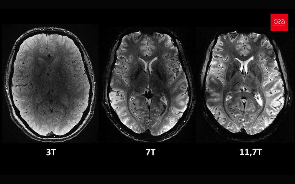
Today, the CEA (the French Alternative Energies and Atomic Energy Commission) unveiled, for the first time worldwide, a series of brain images obtained with the Iseult MRI scanner at a level of resolution never before achieved. Iseult is equipped with an unparalleled magnetic field of 11.7 Tesla, which makes it the world’s most powerful MRI scanner. It aims to help scientists discover new details about the brain’s connections, and activity. DirectIndustry attended the press conference this morning.
From CERN to the Human Brain Microscope
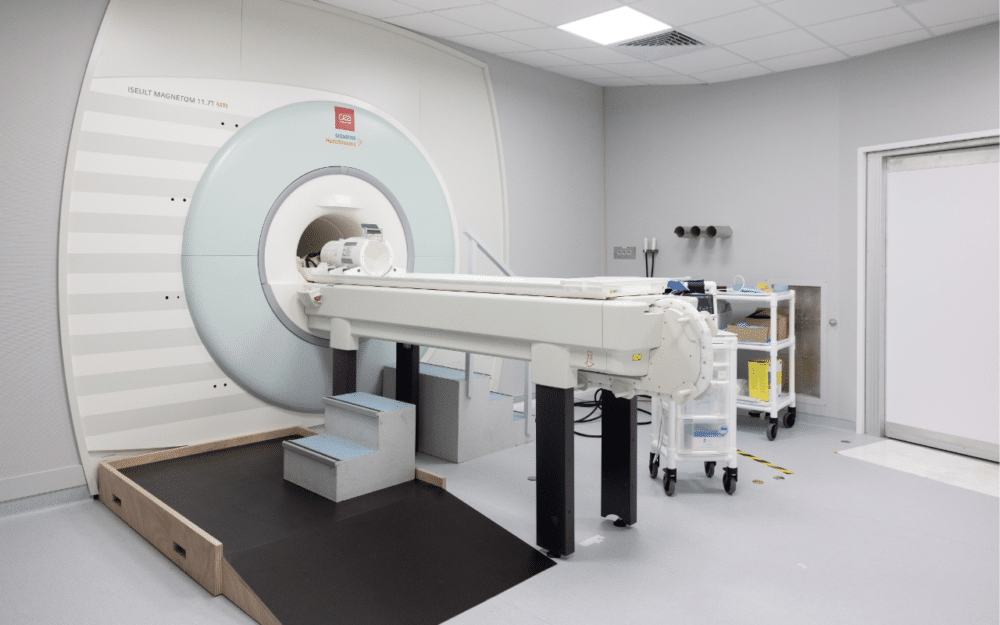
It took the CEA and its partners 25 years to develop this powerful MRI (Magnetic Resonance Imaging) scanner, Iseult. The MRI, which uses an extraordinarily high magnetic field of 11.7 Tesla, weighs 132 tonnes, is 5 meters long, and has a 5-meter diameter. It is powered by 182 kilometers of superconducting wires and sustains 1,500 amperes flowing through the coil. Thanks to all these features, Iseult was able to obtain these unprecedented anatomical images of the human brain from 20 volunteers in just 4 minutes.
A significant breakthrough is the very high resolution of the images, achieved in a short acquisition time: 0.2 mm in-plane and 1 mm in depth, representing a volume equivalent to only a few thousand neurons. For comparison, achieving the same image quality would theoretically require several hours on an MRI scanner typically found in hospitals (1.5 or 3 Tesla), which is unrealistic due to patient comfort and motion-induced image blurring.
By reaching such fine resolutions, it will be possible to access information about neurons that was previously unattainable and to understand how our brain encodes our mental representations, and learning processes, and even discover the neuronal signatures of consciousness states. This true microscope of the human brain could thus help us better understand the human brain and neurological diseases such as Alzheimer’s or Parkinson’s.
Anne-Isabelle Etienvre, Director of Fundamental Research at CEA, discussed the genesis of the project in the 2000s.
“All the skills were brought together in this laboratory, from neuroscientists to bioinformaticians, with highly complementary expertise. The idea was to build an instrument for research, Iseult, with truly extraordinary characteristics. It was innovative, futuristic, and even downright impossible to achieve.”
The study is currently under review by the journal Nature.
Academic and Industrial Partnerships
This technical feat was made possible thanks to several partnerships involved in the conception and construction of superconducting magnets.
The neurobiologists at the heart of the project mobilized the expertise of another laboratory at the CEA, specialized in superconducting magnets. This expertise had been notably utilized in the construction of the Large Hadron Collider (LHC) at CERN. These scientists also contributed to research on fusion, specifically Tokamaks. They were therefore mobilized to design this superconducting magnet with a very high magnetic field.
According to Pierre Védrine, physicist and specialist in superconducting magnets,
“The material of the magnet is niobium-titanium used at a temperature of -271°C. This is a material commonly used in MRIs but which has been pushed to its limits because the magnetic field of 11.7 Tesla is at the limit of what can be achieved with this material.”
The construction of the magnet was then entrusted to the industrial company Alstom, which later became GE, and it was built in France. Siemens Healthineers did the installation of complementary components of the magnetic resonance imaging system. Guerbet, a manufacturer of contrast agents, used the CEA’s high-field MRI platform to evaluate and select molecules with strong potential applications in humans. The University of Freiburg in Germany developed new technologies and methods for ultra-high-field MRI.
Installed at the CEA, the world’s most powerful MRI cost €70 million. €50 million was allocated to the construction of the magnet.
How Does an MRI Work?
For Nicolas Boulant, Project Manager of Iseult and research director at the CEA,
“When we talk about MRI, we refer to “imaging” as we visualize images. “Magnetic” refers to the fact that we visualize the magnetic properties of the atoms that make up our bodies, mainly water. Our bodies are composed of 80% water, so we have a significant signal when observing this. But we need an external magnetic field, provided here by the magnet at the heart of the MRI, to polarize the medium and obtain a measurable macroscopic signal. “Resonance” refers to the electromagnetic waves that excite this matter and tune its frequency to the magnetic field available.”
If today’s announcement is a world first in the field of medical imaging it is due to several acquisitions made on humans in 2023 at an unparalleled magnetic field of 11.7 Tesla, explained Nicolas Boulant:
“The record up to today was 10.5 T in the USA. In the hospital setting, we have MRIs of 1.5 T or even 3 T. 11.7 T is 230,000 times the Earth’s magnetic field.”
Why Increase the Magnetic Field?
In the absence of a magnetic field, the small microscopic magnets associated with atoms point in random directions, it’s complete chaos. When all this is placed within an MRI with a magnetic field, it aligns harmoniously to provide a measurable signal, detailed Mr. Boulant:
“The higher this field, the more amplified the response, and we can monetize this response with greater resolution and clarity in the images. Only Iseult, with its unparalleled magnetic field, can provide image clarity at this mesoscopic resolution.”
In essence, it will be possible to observe neurons in action more effectively. With this strong magnetic field, it will also be possible to observe other less abundant and less detectable chemical species than water, such as phosphorus, fluorine, and sodium, which provide information about the brain’s biochemistry.
Side Effects?
But a stronger magnetic field also means a denser magnetic field. And so the teams had to obtain authorization from the competent health authorities. The study aimed to confirm the absence of side effects on the health of the 20 healthy volunteers who took part in the experiment. This only takes 4 minutes to be compatible with clinical practices.
According to Nicolas Boulant,
“All volunteers are doing well. We have not been able to establish any significant health effects on the volunteers related to exposure to the magnetic field.”
RELATED ARTICLE
Medical Applications
The details obtained with the Iseult MRI will have significant applications in medical research. At a time when the population is aging, the potential offered by this machine to detect diseases at an early stage is of societal importance.
The ultra-fine anatomical information, for example, contributes to establishing a better diagnosis and management of neurodegenerative diseases such as Alzheimer’s or Parkinson’s disease, predicts Mr. Boulant:
“For example, the images could reveal a concentration of iron, which could subsequently inform us about diseases such as Parkinson’s. This would enable the detection of this disease at an earlier stage and the development of new treatments.”
The Iseult MRI will also facilitate the detection of weak signals, underutilized at low field strengths such as that of lithium. This medication is used to treat schizophrenia and bipolar disorders. While it is effective, its action is not well understood.
“Iseult, with its unparalleled sensitivity, will thus allow us to further investigate the distribution of this agent to better understand its role.”
So when will such an MRI be available in a hospital setting? For Nicolas, practical deployment seems challenging:
“Given the complexity and costs involved, operating an MRI with such an unparalleled magnetic field is not simple. The higher the magnetic field, the more complex it is to operate. It requires a certain expertise, a certain know-how that we have acquired here at the CEA. We consider Iseult as a research instrument to acquire fundamental knowledge that we hope to transpose and implement in a hospital setting someday.”
For the moment, the MRI will be used on healthy patients. Pathological patients will have to wait.
The CEA also hopes to capitalize on the future by preparing the next generation of superconducting magnets, particularly regarding the use of new materials.
Key Figures
Magnetic field: 11.7 Tesla (1.5 and 3 T for MRI machines in hospitals)
MRI Specs: 132 tonnes, 5 meters long, 5 meters in diameter
182 kilometers of superconducting wires
1,500 amperes flowing in the coil
Magnet: cooled to -271.35 °C using 7,500 liters of liquid helium
90 cm central bore
5 hours for the current ramp-up


![Image [Best of 2025] Power Moves in the Energy World](/wp-content/uploads/sites/3/energy-320x213.jpg)
![Image [Best of 2025] How Generative AI Is Transforming Industry](/wp-content/uploads/sites/3/AI-4-320x213.jpg)
![Image [BUYING GUIDE] How to Choose the Right Industrial Robot?](/wp-content/uploads/sites/3/Industrial-Robot-320x213.jpg)
![Image [Buying Guide] How to Choose the Right Safety Shoes?](/wp-content/uploads/sites/3/Safety-Shoes-320x213.jpg)