

How to choose the right microscope which helps users achieve efficient workflows? This article discusses factors users should consider before selecting a digital microscope for inspection and quality control (QC) as well as failure analysis (FA) and research and development (R&D).
Content provided by Leica
Understanding well beforehand the application requirements and user needs for industries, like automotive, electronics, machine engineering, and medical devices is key. The microscope solution should help users achieve efficient and reliable inspection, QC, FA, and R&D. It should be easy to operate, adapt to user needs, and make reporting and sharing of results simple.
Why Use Digital Inspection Microscopy?
Today many industries, like automotive, transportation, electronics, machine engineering, and medical devices, use more and more workflow-centric processes. The goal is to manufacture products with better performance and longer lifetimes while still maintaining cost-effective manufacturing which conforms to ever-stricter specifications and standards.
Inspection of components and parts for industrial manufacturing and production, process engineering, quality control, and assurance (QC/QA), failure analysis (FA), product innovation, or research and development (R&D) often is done with the aid of a microscope. The capabilities of the microscope used can make a big difference in terms of inspection efficiency [1,2]. For more details about what to consider when choosing a routine inspection microscope, the reader can refer to reference 1.
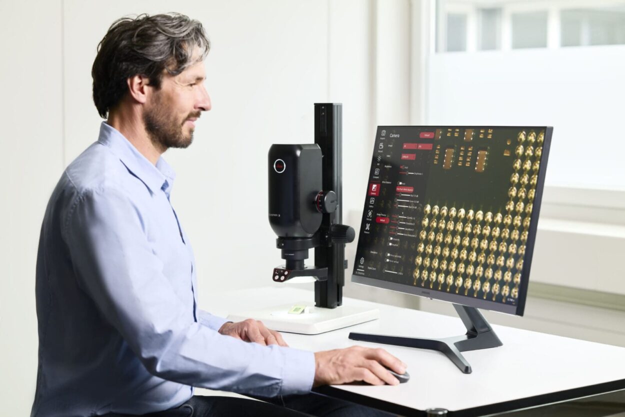
Inspection, documentation, and in-depth analysis of components and parts to determine conformity with product specifications can be done efficiently, reliably, and ergonomically with digital microscopy [2,3]. Digital microscopes do not use eyepieces, but instead, the live image is displayed directly on a monitor.
When deciding upon a digital microscope to utilize for inspection, users should confirm that the microscope offers the optical performance and customizability needed to meet their needs for inspection, QC, FA, and R&D. To help users when selecting a digital microscope for inspection, the main factors to consider are discussed below.
Factors to Consider
Magnification and Resolution
Some components require inspection over a large range of scale: from macroscopic (> 2 mm) to mesoscopic (< 2 mm – 50 µm) and microscopic (< 50 µm – 1 µm) [refer to figure 1].

Concerning digital microscope performance, important factors to consider for inspection over these size scales are [1]:
- High enough magnification and resolution in order to reveal fine details at the mesoscale or microscale
- Zoom range which enables users to quickly go from component overview to seeing its fine details (refer to the example below)
The performance of a microscope is also dependent on optics which are corrected for chromatic aberrations and image flatness, e.g., apochromatic and planarity correction [1].
Main Areas of Application
It is also important to determine the main areas of application for which the digital microscope should be used. These areas concerning inspection and quality control (QC), failure analysis (FA), and research and development (R&D) are described below.
In-Line, At-Line, or Off-Line Quality Control (QC)
In most cases, inspection and in-line and at-line QC are done during production directly at the production site to detect the presence of any defects or irregularities. Quick inspection or screening at strategic points in the production process can help ensure that the standards and specifications are being met. Off-line QC is normally done at various stages of production, but away from the production site, to further minimize or even eliminate defective products which do not conform to the targeted specifications. Off-line QC occurs less rapidly than in-line or at-line QC and tends to require a more detailed investigation of parts or components.
Quick Check or In-Depth Analysis (FA or R&D)
For FA stemming from production or service as well as prototype design and product development (R&D), quick checks and in-depth analyses of parts and components can be required at times. Either quick checks or in-depth analysis can be used for such things as root-cause analysis of failures and prototype investigation when doing R&D. Root-cause analysis can often require a detailed evaluation of 1 or more components or connections to understand clearly what causes the product to fail. During product development, often prototype designs are optimized with the help of quick checks or in-depth analyses of components and connections to verify product performance and the ability to achieve an efficient method of production.
Leica digital microscopes for efficient inspection
Area of appropriate application
The different inspection, QC, FA, and R&D needs of users can be addressed with the appropriate Leica digital microscope (refer to figure 2 and table 1 below):
- The Emspira 3 digital microscope enables efficient inspection, basic analysis, and documentation for in-line or at-line QC, and quick checks for FA and R&D at the macroscale to mesoscale (> 2 mm – 50 µm)
- The DVM6 digital microscope permits efficient inspection, detailed analysis, and documentation, for off-line QC and in-depth analysis for FA and R&D at the mesoscale to microscale (< 2 mm – 1 µm)

Application Areas of Leica Digital Microscopes
| In-line or At-line QC / Quick checks for FA or R&D | Off-line QC / In-depth analyses for FA or R&D |
| Emspira 3 | DVM6 |
| Macro- to mesoscale (> 2 mm – 50 µm) | Meso- to microscale (< 2 mm – 1 µm) |
Advantages of Leica Digital Microscopes
A comparison of the advantages for users when using the Emspira 3 or DVM6 digital microscope is shown in table 2 below.
| Emspira 3 | DVM6 |
| See a large area of the component in a single image with a large overview (maximum area = 76 mm x 43 mm) | Easy storage and rapid recall of important parameters, such as, optics, illumination, tilting angle, and camera settings, with an encoded system |
| Zoom in quickly onto an area of interest to reveal details at higher magnification and resolution | Find the component details quickly and easily using the hybrid XYZ stage; it can be moved and rotated both manually and with motorized control |
| Easy storage and rapid recall of important parameters, such as optics, illumination, tilting angle, and camera settings, with an encoded system | Visualize hard-to-reveal details of components rapidly with tilting stand and integrated versatile contrast methods, e.g., LED ring light and coaxial illumination; fully integrated so no need to install them during use |
| Ensure correct results with encoded zoom optics | Obtain quickly large mosaic overviews of areas with large height differences using extend depth of field (EDoF) and XYZ stitching |
| Work with confidence in industrial environments as robust IP 21 housing protects internal optics and mechanics | Obtain quickly large mosaic overviews of areas with large height differences using extended depth of field (EDoF) and XYZ stitching |
| Reduced risk of spreading germs thanks to the antimicrobial surface | Just start investigations by taking advantage of the autofocus keeping the image in focus while navigating over the sample |
| Maximum resolution of 3 µm (337 line pairs/mm) | Maximum resolution of 0.42 µm (2,366 line pairs/mm) |
| Acquire sharp images with a 12-megapixel camera sensor | Acquire sharp images with a 10-megapixel camera sensor |
Example for In-Line, At-Line, or Off-Line Qc: Inspection of an Electronic Device
In-Line or At-Line QC
When doing an inspection for defects or errors during in-line QC, usually a microscope is used to:
- Obtain a large overview of the component at lower magnification.
- Rapidly zoom in on an interesting area of the component which requires more detailed inspection at higher magnification to see the fine details.
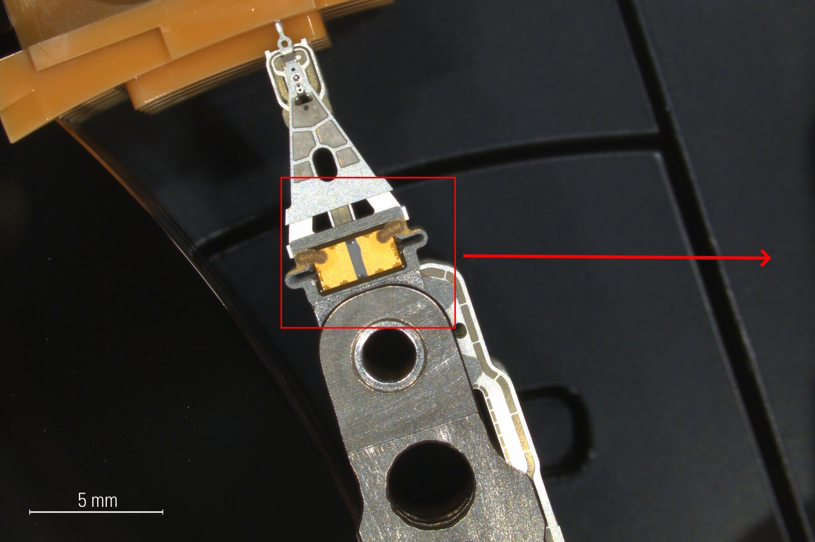
As a possible example of in-line or at-line QC, figure 3 below shows images of hard drive components recorded with a Leica digital microscope like the Emspira 3. For this case, an overview of the disk or platter read-write head and actuator arm (figure 3a) of a hard drive is seen. Then, by easily and quickly increasing the zoom factor, an image of the read-write head and actuator arm (figure 3b) showing scratches (defects) at higher magnification can be recorded for documentation.


Off-line QC
For inspection during off-line QC, microscopes often can be used to make a more detailed examination of parts and components which is not practical or possible for in-line QC. As a possible example of off-line QC, figure 4 below shows a portion of the underside of a hard drive which is the bottom of a PCB. The images were taken with the DVM6 microscope using the integrated ring light (figure 4a) and coaxial illumination with the quarter wave plate and relief contrast (figure 4b). The pads, traces, through-holes, and substrate surface are seen. Different details of the hard-drive PCB bottom, such as scratches, defects, and contamination, are better enhanced and more noticeable in one image than the other. The integrated illumination and multiple contrast methods of the DVM6 enable users to visualize and document difficult-to-see fine details of components, as shown here with the hard-drive PCB example, in a more efficient way, as there is no need to change the setup of the microscope.


Summary
Many industries have an ever-growing demand for faster and lower-cost production of parts and components while meeting ever-stricter product specifications. As a result, manufacturers are making their workflows more and more efficient, whether for inspection and production, quality control and assurance (QC/QA), failure analysis (FA), or research and development (R&D). Normally, the workflow stretches from the macroscale through the mesoscale to the microscale.
Leica digital microscopes, which operate without eyepieces, and the live image of the part or component is observed directly on a monitor, enable users to work efficiently and ergonomically. They can be used for inspection, QC/QA, FA, and R&D in various industries to optimize the entire workflow. The factors to consider when choosing the most appropriate digital microscope for users’ needs were described in this article.
References
- J. DeRose, D. Barbero, How to select the right solution for visual inspection: Factors to consider when looking for a routine inspection microscope, Science Lab (2021) Leica Microsystems.
- J. DeRose, G. Schlaffer, What You Always Wanted to Know About Digital Microscopy, but Never Got Around to Asking, Science Lab (2015) Leica Microsystems.
- J. DeRose, G. Schlaffer, Digital Microscopy with Versatile Illumination and Various Contrast Methods for More Efficient Inspection and Quality Control: Example applications using the Leica DVM6 with integrated ring light or coaxial illumination system, Science Lab (2017) Leica Microsystems.


